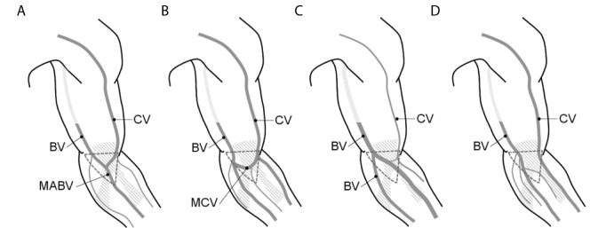
Figure 3.
The superficial veins of the upper limb present a certain inter-individual variability in their running pattern and caliber; A) in type I, cephalic vein (CV) and basilic vein (BV) merge into the median antebrachial vein (MABV) of the forearm; B) in type II, the median cubital vein (MCV) forms an anastomose between CV and BV; C) in type III, CV is threadlike and BV splits in two branches of the forearm; D) in type IV, CV and BV run in parallel with no evident superficial anastomoses; the dashed triangle delimits the cubital fossa area.

