
Fig. 7. In monolayer culture, cellular morphology differs from in vivo morphology.
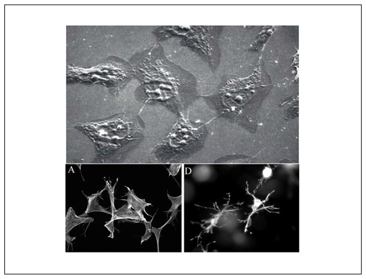
Upper picture: Fibroblasts in monolayer culture on lamella observed by scanning electron microscopy (SEM): population sub-confluence. Cells have rather starry shapes. Length of the line 10 μm (Image: A. Minondo, L’Oréal); lower pictures: human fibroblasts project a dendritic network of extensions in collagen matrices but not on collagen-coated coverslips. Fibroblasts were incubated for 5 h on collagen-coated surfaces (A) or in collagen matrices (D). At the end of the incubations, samples were fixed and stained for actin. Reprinted with permission from Grinnell et al. (2003).