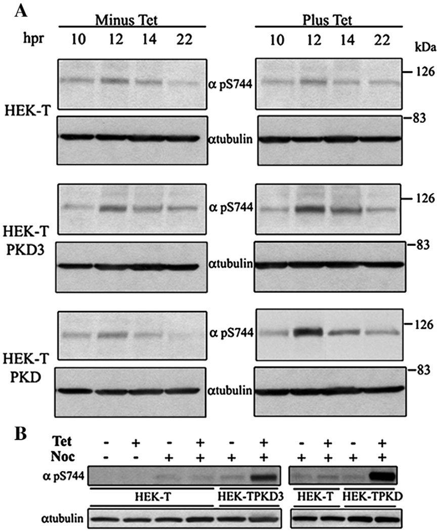
Fig. 2.
PKD3 Ser731 and PKD Ser744 phosphorylation during mitosis. HEK-T (control), HEK-TPKD3 and HEK-TPKD cells, without or with Tet (100 ng/ml) synchronized at G1/S by aphidicolin treatment (A), were collected at the indicated times after aphidicolin removal. Alternatively, the cultures, without or with Tet (100 ng/ml), were synchronized at G2/M by nocodazole treatment (B) for 12 h. Equal amounts of cell lysates obtained from aphidicolin or nocodazole-treated cells were resolved in a 4.5–15% SDS-PAGE. Proteins were transferred to PVDF membranes and the membranes incubated with pS744 or α-tubulin antibodies. Signals were detected as described under Materials and methods.

