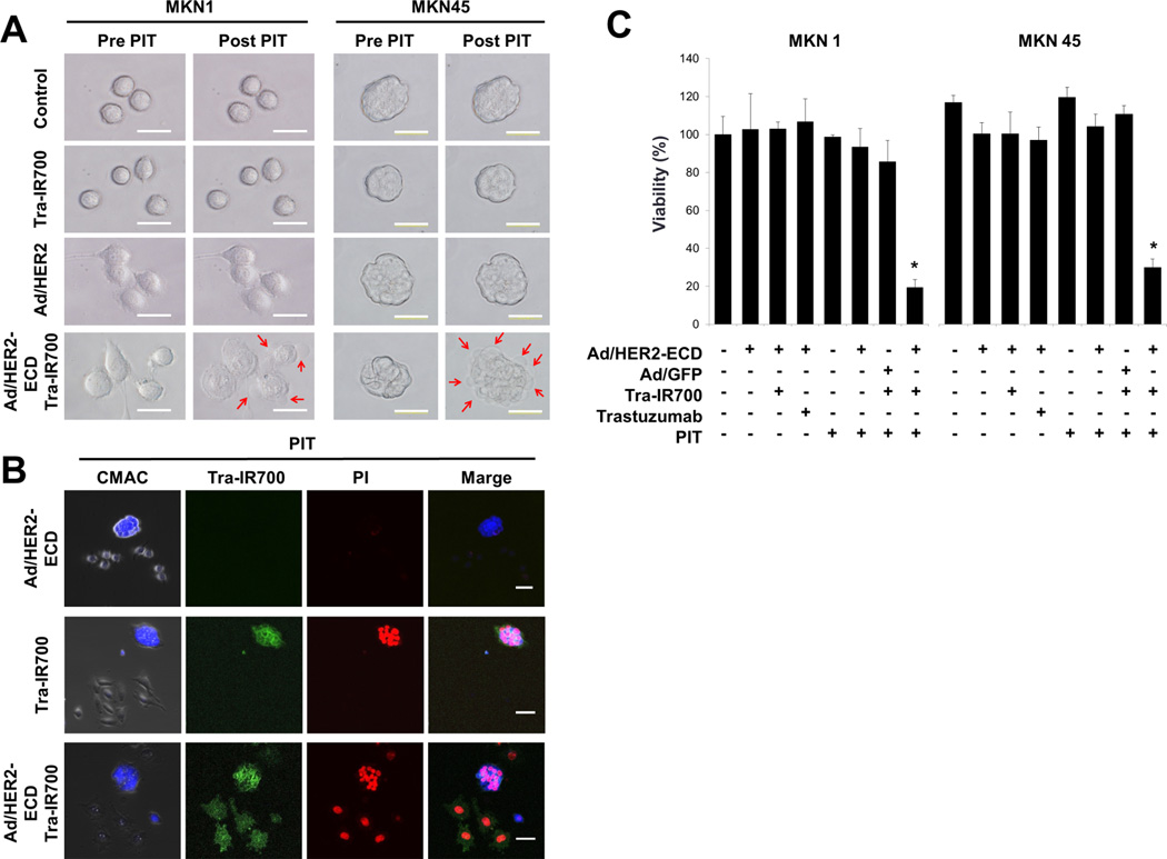
Figure 2.
Microscopic analysis of the HER2-ECD-induced HER2-negative cells treated with Tra-IR700-mediated PIT. A, morphological changes of the tumor cells after PIT. MKN1 and MKN45 were cultured with Ad/HER2-ECD for 48 h and were treated with Tra-IR700 for 6 h. Changes in microscopic morphology were observed before and after PIT with 5 J/cm2 at 690 nm. Treated cells were morphologically captured as swelling like a balloon after PIT. Bleb formations on cellular membranes are indicated with red arrows. B, Tra-IR700 mediated PIT-induced cell death. N87 cells were labeled with Cell Tracker® Blue CMAC dye and then co-cultured with unstained MKN1 cells. These cells were exposed to Tra-IR700 for 1 h and were irradiated with near infrared light at 5 J/cm2. After irradiation, the cells were stained with propidium iodide (PI) for identifying dead cells. Immunohistochemistry showed that PI stained only labeled N87 cells bound by Tra-IR700 (middle row). The co-cultured plate was infected with Ad/HER2-ECD at an MOI of 50 for 48 h and treated with Tra-IR700-mediated PIT. PI stained labeled N87 cells as well as unlabeled MKN1 cells (bottom row). C, a comparison of the cell viability after the treatment of the Ad/HER2-ECD with Tra-IR700-mediated PIT. MKN1 and MKN45 cells were seeded on 96-well plates (n= 4 per group) for 24 h. Ad/HER2-ECD were infected at an MOI of 50 for 48 h. After 24 h of treatment with trastuzumab and Tra-IR700, the cells were irradiated with NIR light (10 J/cm2). An XTT assay demonstrated the statistically significant loss of cell viability in only the integration of Ad/HER2-ECD to the Tra-IR700-mediated PIT group as compared to other groups (n= 4, * Cell viability, P status: MKN1; 19%, P< 0.01, MKN45; 26%, P< 0.01, using the Student’s t test).

