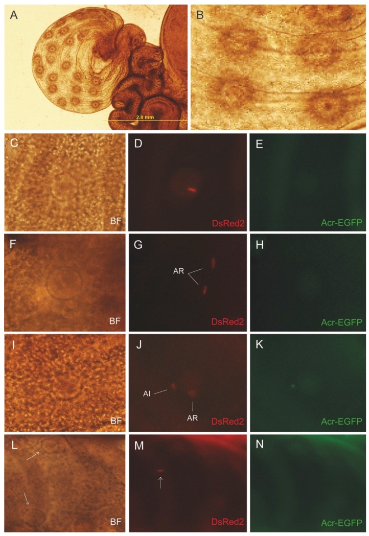
Figure 5.
Most of the sperm in the ampulla underwent acrosomal exocytosis. A) Representative image of an ampulla after ovulation. B) The eggs were easily observed through the oviductal wall. C-E) Representative images of a fertilized egg where it was possible to observe the fluorescence of the tail. F-H) Representative images of two sperm in the vicinity of an egg. Both sperm lost their acrosomes. I-K) Representative images of two sperm in the vicinity of an egg. In this case, one sperm was acrosome intact (AI) while the other already underwent acrosomal exocytosis (AR). L-N) Representative example of one acrosome reacted sperm swimming through 3 ovulated eggs (indicated by arrows). BF: bright field image,

