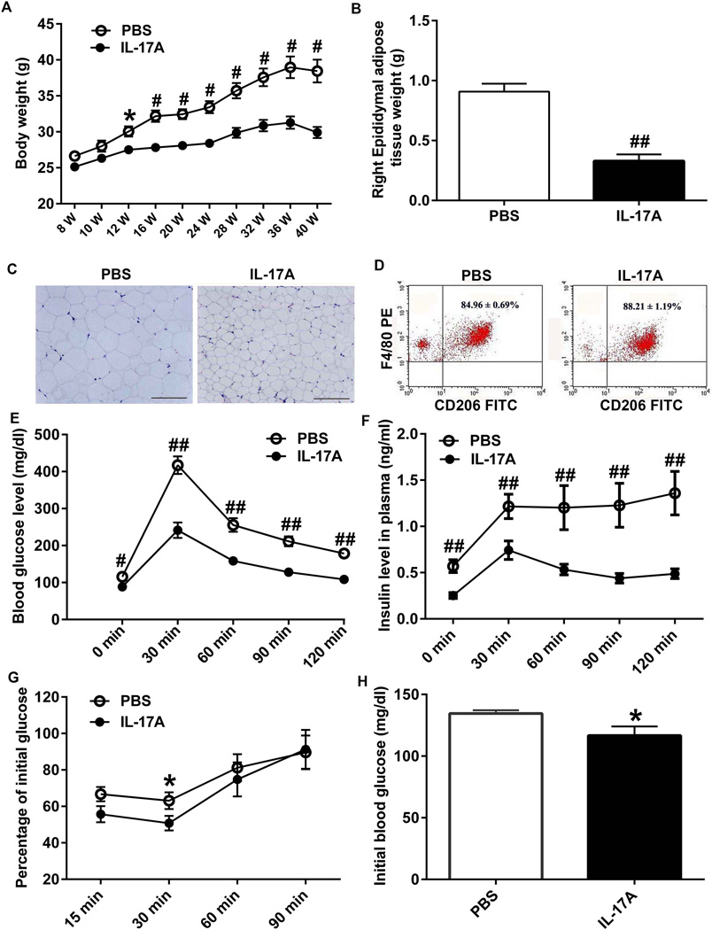
Figure 7. Effect of IL-17A overexpression on bodyweight, IPGTT, ITT and adipose tissue.
(A) Two-month-old mice (n=9–11/each group) were subjected to intraventricular injection of PBS or rAAV5-IL-17A and their bodyweights were measured every two weeks. (B) Thirty-two weeks after the injection, mice were euthanized and epididymal adipose tissues were isolated. The average weight of right epididymal adipose tissues in IL-17A-in-Brain mice are approximately one third of that in PBS-in-Brain mice. (C) Haematoxylin and eosin-stained images of paraffin embedded epididymal adipose tissue sections from PBS-in-Brain and IL-17A-in-Brain mice. Scale bars are 200µm. (D) Flow cytometer analyses showing expression levels of CD206+ and F4/80+ M2 macrophages in the adipose tissues. (E and F) Twenty weeks after injection, mice were fasted for 16 h and injected i.p. with 1.5 g glucose/kg bodyweight and blood glucose and insulin were measured before and after glucose injection at the indicated time points. (G) Thirty weeks after PBS or rAAV5-IL-17A injection, mice were fasted for 6 h and injected i.p. with 0.75 U insulin/kg bodyweight for ITT. Blood glucose levels were determined and are shown as percent changes from (H) basal blood glucose levels. * P < 0.05, # P < 0.01 and ## P < 0.001.

