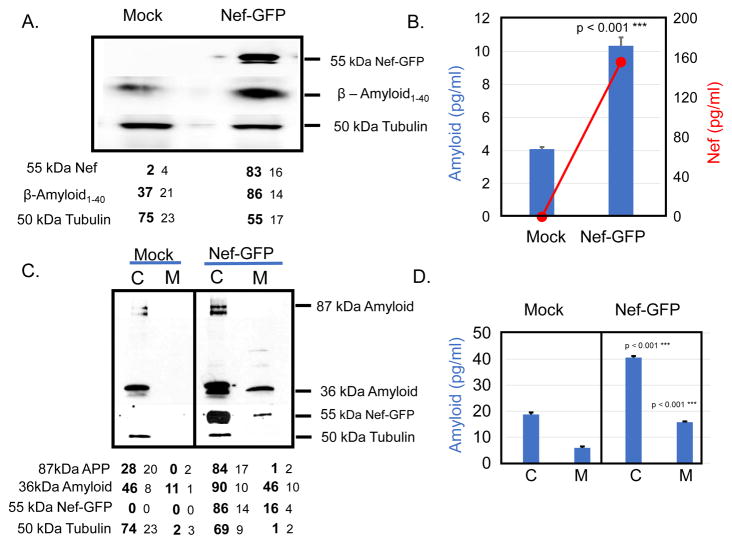
Fig. 2.
Transfection of SH-SY5Y cells with a Nef-GFP expression plasmid or treatment with Nef exosomes increases the amount of Aβ produced in target cells. a SH-SY5Y cells were transfected with 30μg of Nef-GFP expression plasmid or mock transfected with GFP vector. After 48 h cells were harvested and immunoblot was used to detect the amounts of Nef-GFP and Aβ1-40. b ELISA was also run to determine the amount of Nef-GFP (red line, red axis) and Aβ1-42 (blue bars, blue axis). c SH-SY5Y cells were treated with Nef exosomes isolated from transfected HEK293 cells or mock treated cells. 24 hours after treatment, the cells were harvested and lysed (P for cell pellet). We also collected conditioned medium and subjected it to ultra-centrifugation at 100,000 x g (S for supernatant) and run on a PAGE gel. The gel was blotted with antibody that reacts with intermediate forms of Aβ (see 36 kDa band and higher molecular weight forms). The blot was stripped and re-probed with antibody against the 87kDa APP, Nef-GFP and Tubulin. The numbers below the gel are the average and SD of three independent experiments. d The cell pellet (C) and conditioned medium (M) were also subjected to ELISA to quantitate Aβ1-42. The results shown are from three independent experiments.

