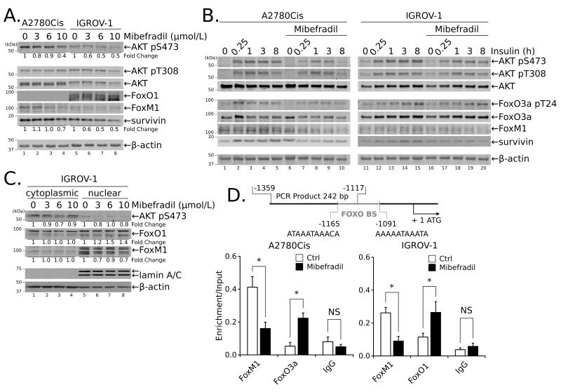
Figure 3.
Treatment with Mib reduces survivin expression in ovarian cancer cells through disruption of PI3K/AKT and Forkhead box protein activities and sub-cellular localization. (A) A2780Cis and IGROV-1 cells were treated with the indicated concentrations of Mib for 24 hours prior to blotting for AKT phosphorylation and FoxO1 and FoxM1 protein expression. (B) Cells were treated with 10 μmol/L Mib for 1 hour followed by stimulation with 10 μg/ml insulin for up to 8 hours. (C) Subcellular localization of FoxO1 and FoxM1 was determined by blotting cytoplasmic or nuclear extracts of IGROV-1 cells treated with increasing concentrations of Mib for 24 hours. (D) A2780Cis cells or IGROV cells were treated with Mib (10 μmol/L) or DMSO for 24 hours, cross-linked and subjected to ChIP using antibodies specific for the indicated proteins; rabbit IgG served as a negative control. Enrichment in binding of specific proteins was determined based on the amplification of immunoprecipitated DNA by qPCR of the BIRC5 promoter using specific primers spanning the FOXO binding sites (FOXO BS; schematic on top). The data were normalized to the corresponding input DNA as described (see Materials and Methods) and are presented as the mean from two independent experiments ± SEM; *, P < 0.05, **, P < 0.01.

