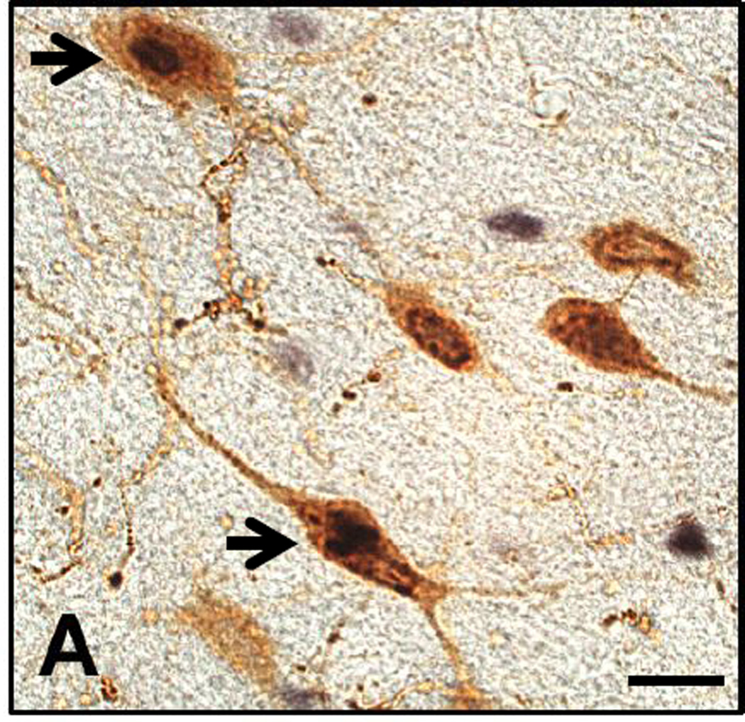
Figure 1.
High-power photomicrograph of coronal section through the lateral hypothalamus immunolabelled for Fos-protein (black nuclear stain) and orexin-A (brown cytoplasmic stain). Arrows indicate co-labeled neurons. Other orexin neurons without Fos staining and Fos staining in non-orexin neurons are shown. Scale bar = 20 µm.

