Figure 1.

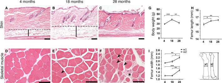
Age‐related degenerative changes in wild‐type mice. Representative hematoxylin and eosin staining of skin (A–C) and skeletal muscle (D–F). (A–B) Increased initial thickening of the hypodermal fat layer in 18‐month‐old mice (B) becomes virtually absent in 28‐month‐old mice with terminal marked dermal fibrosis (C). (D–F) Increased degenerative changes with age in muscle. (D) 4‐month‐old mice showed essentially no degenerative changes. (E) 18‐month‐old mice have mild variation in fiber size and rare centralization of the nuclei (arrowhead). (F) 28‐month‐old animals present marked variable fiber size, frequent nuclear centralization (arrowheads), and endomysial fibrosis (star). (G) A significant increase in body weight between the 4‐ and 18‐month‐old mice decreased at 28 months. (H) Femur lengths with initial increase and posterior plateau phase. (I) Femur thickness measured in the mid‐diaphysis of 2 bone dimensions (w1 and w2). Dashed line (hypodermal mean upper limit). Vertical bars (hypodermis). Scale bars 100 μm. *P < 0.05, **P < 0.01 and ***P < 0.001; one‐tailed paired t‐test, the data are expressed as the means ± SEM.
