
Figure 2.
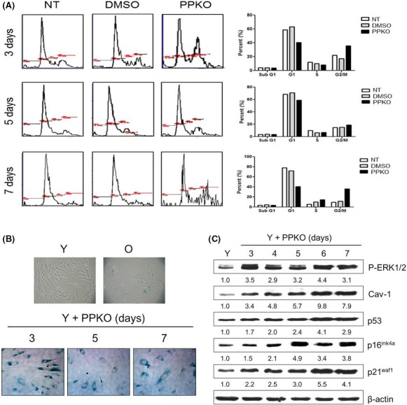
G2/M cell cycle arrest and senescence‐like changes induced by PPKO in young HDFs. A. Cell cycle analysis. B. SA‐β‐gal staining. C. Induction of senescence‐associated proteins in ketoxime‐treated young HDFs. (A) Young HDFs were cultured for 1 day and serum‐depleted by incubation with SFM for 1 day. The cells were then stimulated with 10% FBS in the presence of vehicle or 1 mM PPKO for 3–7 days. The cells were stained with PI, and the cell cycle was analyzed by flow cytometry. The percentage of cells at sub‐G1, Go/G1, S, and G2/M was plotted and shown on the right side. (B) Young HDFs were treated with vehicle (Y) or 1 mM PPKO for the indicated times (3–7 days). Old HDFs (O) were also treated with vehicle for control purposes. Cells were then stained with X‐gal to monitor SA‐β‐gal activities and photographed under an inverted microscope at 100×. (C) Young HDFs were treated with vehicle (Y) or 1 mM PPKO for 3–7 days, and cell lysates were then analyzed by Western blot analysis using antibodies against senescence‐associated proteins (P‐ERK1/2, Cav‐1, p53, p16ink4a, and p21waf1). β‐actin was used as an internal control. Bands in blots were normalized to β‐actin in each lane. Fold increases vs. levels in lane Y are written under each band.