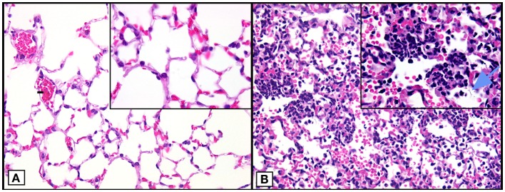
Figure 1.
Histopathology of mouse lungs in normal lungs and lungs after ALI following airway deposition of IgGIC in C57BL/6J mice. (A) Histological features of normal mouse lung. (B) Positive control (acutely injured lung induced by airway deposition of IgGIC) 6 h after induction of ALI showing lung infiltration of PMNs, intra-alveolar hemorrhage, and hyaline membranes (arrow). Images modified from Bosmann et al. (18). Both frames are from paraffin-embedded sections stained with hematoxylin and eosin (×40 view; insets: ×100 view).

