
Figure 1.
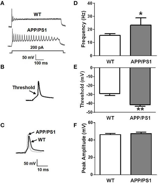
Increased neuronal excitability in APP/PS1 mice. (A) Representative recording traces of action potential repetitive firing. (B) The schematic of threshold. (C) Representative recording traces of action potential peak amplitude. (D) The mean number of APs elicited by 200 pA depolarizing current. Neurons from APP/PS1 mice (n = 10) fired significantly more APs than WT (n = 12). (E) AP threshold in APP/PS1 mice cultured pyramidal neurons was significantly lower than WT mice (n = 9 per group). (F) The peak amplitude of APs in WT and APP/PS1 (n = 9 per group) mice cultured pyramidal neurons didn't show significantly difference. Mean ± SEM was displayed. *Presents p < 0.05; **presents p < 0.01.