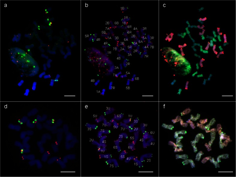
Fig. 2.
Fluorescence in situ hybridization (FISH) using 5S and 25S rDNA (a, d); pAs1 and pSc119.2 (b, e) repetitive DNA probes, and genomic in situ hybridization (GISH) on mitotic chromosomes of triticale (× Triticosecale Wittm.) ‘Lamberto’ (a, b, c) and Ae. variabilis Eig. (d, e, f). On the GISH images: c the R-genome is visualized in red, the A-genome in green and the B-genome in blue; f the Uv-genome is visualized in red and the Sv-genome in green. Scale bars: 10 μm

