. 2016 Feb 29;59(2):137–142. doi: 10.3340/jkns.2016.59.2.137

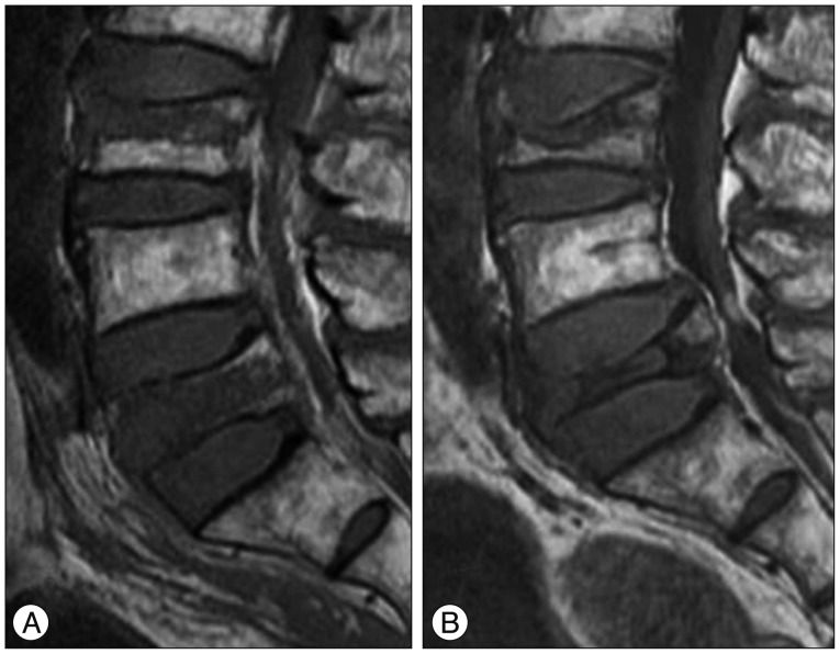
Fig. 3. A 77-year-old man with OVCFs at the L3 and L5 vertebral bodies. A : Sagittal T1WI of initial MRI shows band-like low signal intensity (type 3 pattern) of VCF at the L3 vertebral body and diffuse low signal intensity (type 1 pattern) of VCF at the L5 vertebral body. B : Sagittal T1WI of follow up MRI taken after 4 months shows anterior wedge deformity of VCF at the L3 vertebral body and crush deformity of VCF at the L5 vertebral body. L5 vertebral body exhibits severe degree vertebral body height loss (>40%) and posterior bulging with spinal stenosis. He failed conservative management and underwent surgical treatment (laminectomy with posterior instrumentation). OVCFs : osteoporotic vertebral compression fractures, VCF : vertebral compression fracture.

Fig. 3