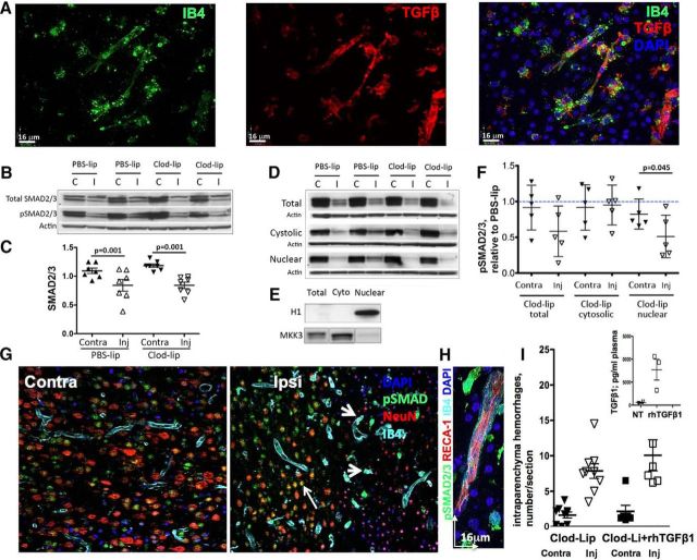
Figure 6.
Replenishment of peripheral TGFβ1 does not attenuate hemorrhage occurrence at 24 h after tMCAO in rat pups with depleted microglia. A, Representative images showing expression of TGFβ1 in IB4+ brain vessels and microglia/macrophages in the injured cortex. B, Western blots showing protein expression of SMAD2/3 (top) and pSMAD2/3 (middle) in the same total tissue lysates at 24 h after tMCAO. The data are normalized to actin (bottom) in individual samples. C, tMCAO significantly and similarly reduces SMAD2/3 expression in injured regions of PBS-lip-treated and Clod-lip-treated rats. A common tissue sample was used in multiple Western blots to allow data analysis. D, Western blots showing SMAD2/3 phosphorylation in total lysates and in cytosolic and nuclear fractions. E, Purity of the subcellular fractions is assessed by the presence of specific cytosolic (MKK3) and nuclear (histone H1) proteins. F, Densitometric quantification of pSMAD2/3 protein in total lysates and in the cytosolic and nuclear fractions. Shown are pSMAD2/3 values in individual Clod-lip rats expressed relative to averaged pSMAD2/3 value in PBS-lip rats in the same individual blots. G, Cell-type specific patterns of SMAD2/3 phosphorylation in contralateral (left) and ipsilateral (right) regions. Note SMAD2/3 phosphorylation in NeuN+ neurons (red) in uninjured regions and in the penumbra but not in the ischemic core. pSMAD2/3 is expressed in IB4+ vessels in both uninjured and injured regions. H, High-magnification image of pSMAD2/3 in a IB4+ vessel and in both IB4+ and IB4− cells in the vicinity of a vessel. I, Quantification of the total number of intraparenchymal hemorrhages in rats treated with Clod-lip, with or without intravenous administration of rhTGFβ1. G, Inset, Plasma levels of TGFβ1 measured by Bioplex ELISA at 24 h after tMCAO in untreated rats and in rats injected rhTGFβ1 intravenously 18 h after tMCAO.

