Figure 7.
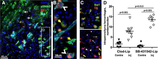
Selective inhibition of TGFbr2/ALK5 signaling in microglial cells induces hemorrhagic transformation 24 h after tMCAO. A, SMAD2/3 phosphorylation is reduced in IB4+ microglia that uptake DiI+/SB-431542-lip delivered intracortically. Essentially all DiI+ cells are IB4+ microglia, and SMAD2/3 phosphorylation is observed in neurons outside of injury, as indicated by thin white arrows. B, Magnified image from a region in A (white box). Yellow arrows point to cells with high pSMAD2/3 expression. White arrowheads point at IB4+/DiI+ cells with low pSMAD2/3 signal. C, Representative example of DiI+/Iba1+/DAPI+ immunofluorescence in the penumbra region of DiI+/SB-431542-lip-treated rat. Essentially all DiI+ cells are double-labeled with Iba1+ (yellow arrows), whereas DiI+/Iba1− cells are infrequent, with low level of label incorporation (arrowheads). There is no DiI+ signal around morphologically integrant nuclei outside of injured region (*, healthy neurons) or nuclei with apoptotic appearance (thin arrows). D, The number of hemorrhages in injured hemisphere is significantly increased in rats treated with SB-431542-lip compared with Clod-lip-treated or nontreated rats.

