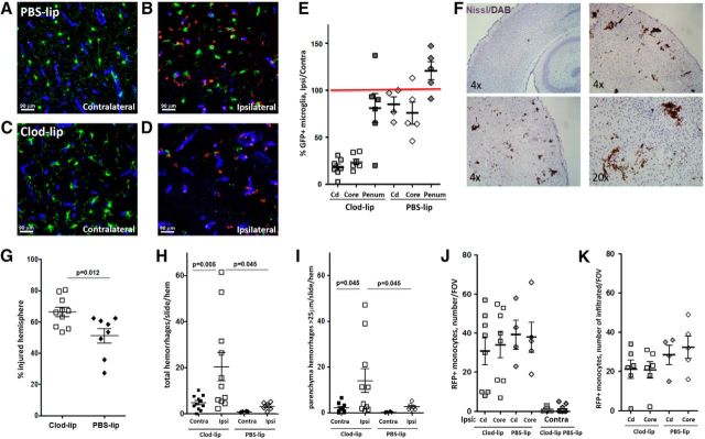
Figure 8.
Microglial depletion exacerbates injury and promotes hemorrhages 24 h following neonatal arterial stroke in P9 mice. A–D, Representative images of Cx3cr1GFP/+ microglia and CCR2RFP/+ monocytes in ischemic core (B, D) and in matching contralateral cortical regions (A, C) of PBS-lip-treated (A, B) and Clod-lip-treated (C, D) mice. Note morphological changes of Cx3cr1GFP/+ in injured regions. Essentially no RFP/+ monocytes are seen in contralateral hemisphere, whereas RFP/+ monocytes are observed both in association with vessels and within injured cortex. E, Quantification of microglial depletion by Clod-lip in the caudate (Cd), the ischemic core, and in the penumbra (Penum). Shown data are percentage of Cx3cr1GFP/+ microglial number in ipsilateral compared with matching contralateral regions in same individual animals. The number of Cx3cr1GFP/+ cells in contralateral hemisphere of Clod-lip-treated mice is lower than in contralateral hemisphere of PBS-lip-treated mice (data not shown). F, Representative examples of hemorrhages identified on DAB/Nissl-stained coronal sections. G, Microglial depletion significantly increases injury volume. Shown are data for individual mice. H, I, Microglial depletion significantly increases the total number of hemorrhages in multiple regions of injured hemisphere (H) and the number of intraparenchyma hemorrhages >25 μm in the longest dimension (I). J, K, The total number of CCR2RFP/+ monocytes (J) and the number of infiltrated CCR2RFP/+ monocytes (K) in individual injured regions are not significantly different between Clod-lip and PBS-lip mice.

