Figure 2.
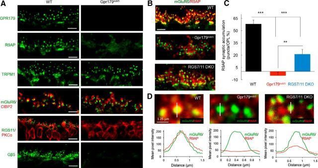
GPR179 is necessary for the correct localization of R9AP in retinal ON-BC synapses and the expression of RGS11 in the OPL. A, Immunohistochemical analysis of ON-BC synaptic components in the OPL of WT and Gpr179nob5 mouse retinas. Retina sections were immunostained with the indicated antibodies. Synaptic architecture in Gpr179nob5 retinas is intact as evidenced by normal apposition of presynaptic and postsynaptic markers (CtBP2, mGluR6, and TRPM1, respectively). Note the lack of accumulation of R9AP and complete absence of RGS11 in Gpr179nob5 ON-BC dendritic tips. Scale bar, 5 μm. B, Double immunolabeling of WT, Gpr179nob5, and RGS7/RGS11 DKO retinas with mGluR6 and R9AP antibodies. Scale bar, 5 μm. Quantification of the accumulation of R9AP within mGluR6-positive puncta of ON-BC dendrites in OPL (described in Materials and Methods). C, Corresponding values. Error bars indicate SEM. *p < 0.05 (one-way ANOVA with Bonferroni's post hoc test). **p < 0.01 (one-way ANOVA with Bonferroni's post hoc test). ***p < 0.001 (one-way ANOVA with Bonferroni's post hoc test). n = 25–30 puncta for each group. D, Top, High-magnification images of retina sections with scan-line analyses of R9AP fluorescence intensity across the midline of mGluR6-positive synaptic puncta in OPL. Bottom, Averaged traces of R9AP fluorescent intensity across the midline of mGluR6-positive synaptic puncta reveal a complete lack of after synaptic R9AP accumulation in Gpr179nob5 retinas, but only a partial loss in RGS7/11 DKO. n = 25–30 for all groups. Scale bar, 0.125 μm.

