Figure 3.

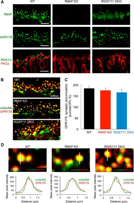
RGS and R9AP proteins are not required for the expression or correct localization of GPR179 in the retinal OPL. A, Immunohistochemical analysis of ON-BC synaptic components in the OPL of WT, R9AP, and RGS7/RGS11 DKO mouse retinas. Retinal sections were stained with the indicated antibodies. Scale bar, 5 μm. B, Double immunolabeling with mGluR6 and GPR179 antibodies. Scale bar, 5 μm. Quantification of the accumulation of GPR179 within mGluR6-positive synaptic puncta relative to the OPL (described in Materials and Methods). C, Corresponding values. Error bars indicate SEM; n = 30–35 puncta for each group. One-way ANOVA shows no significant differences. D, Top, High-magnification images of retinal sections with line-scan analyses of GPR179 fluorescence intensity across the midline of mGluR6-positive synaptic puncta in OPL. Bottom, Averaged traces of GPR179 fluorescent intensity across the midline of mGluR6-positive synaptic puncta reveal no changes to postsynaptic GPR179 accumulation in either R9AP KO or RGS7/RGS11 DKO retinas; n = 25–30 for all groups.
