
Figure 1.
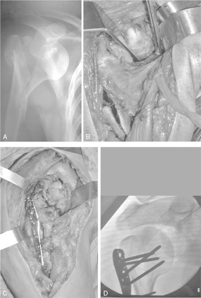
A) Radiograph showing fracture into three parts with anterior dislocation. (B and C) Intraoperative appearance of the fracture and dislocation after reduction and osteosynthesis with a Philos plate. D) Intraoperative fluoroscopic image.
Official websites use .gov
A
.gov website belongs to an official
government organization in the United States.
Secure .gov websites use HTTPS
A lock (
) or https:// means you've safely
connected to the .gov website. Share sensitive
information only on official, secure websites.
A) Radiograph showing fracture into three parts with anterior dislocation. (B and C) Intraoperative appearance of the fracture and dislocation after reduction and osteosynthesis with a Philos plate. D) Intraoperative fluoroscopic image.