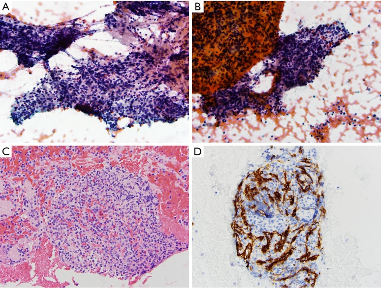
Figure 1.
Case 1: aspirate smears and cell block material. (A&B) Aspirate smears show a polymorphic population of lymphocytes with admixed eosinophils and histiocytes (Pap stain, 400×); (C) cell block section demonstrates similar findings to the aspirate smears (H&E stain, 400×); (D) CD8 stain highlights sinusoidal endothelial cells (CD8 immunohistochemical stain, 400×).

