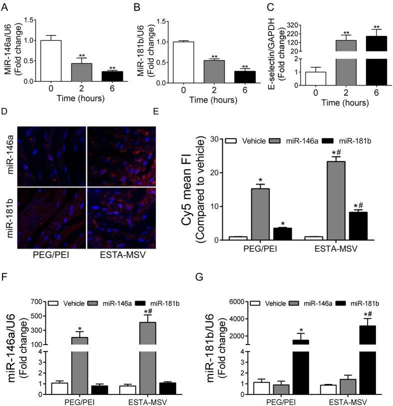
Figure 1. The expression of miR-146a and miR-181b in inflamed endothelial cells and the transfection efficiency of particles.
The expression of miR-146a (A), miR-181b (B), and E-selectin (C) in HMVECs after treatment with TNF-α (10 ng/mL) for 0, 2, and 6 hours. **P < 0.01 vs. 0h. (D) Representative fluorescence images of TNF-α-treated en face mouse aortas transfected with PEG/PEI nanoparticles or ESTA-MSV microparticles containing Cy5-labeled miR-146a and miR-181b. Magnification: 40X. (E) Quantification of Cy5 mean fluorescence intensity. The expression of miR-146a (F) and miR-181b (G) in HMVECs after transfected with the particles as above. Data are shown as the means ± SEM and are representative of 3 independent experiments. *P < 0.05 vs. vehicle groups of the same particle; #P < 0.05 vs. PEG/PEI groups of the same miR.

