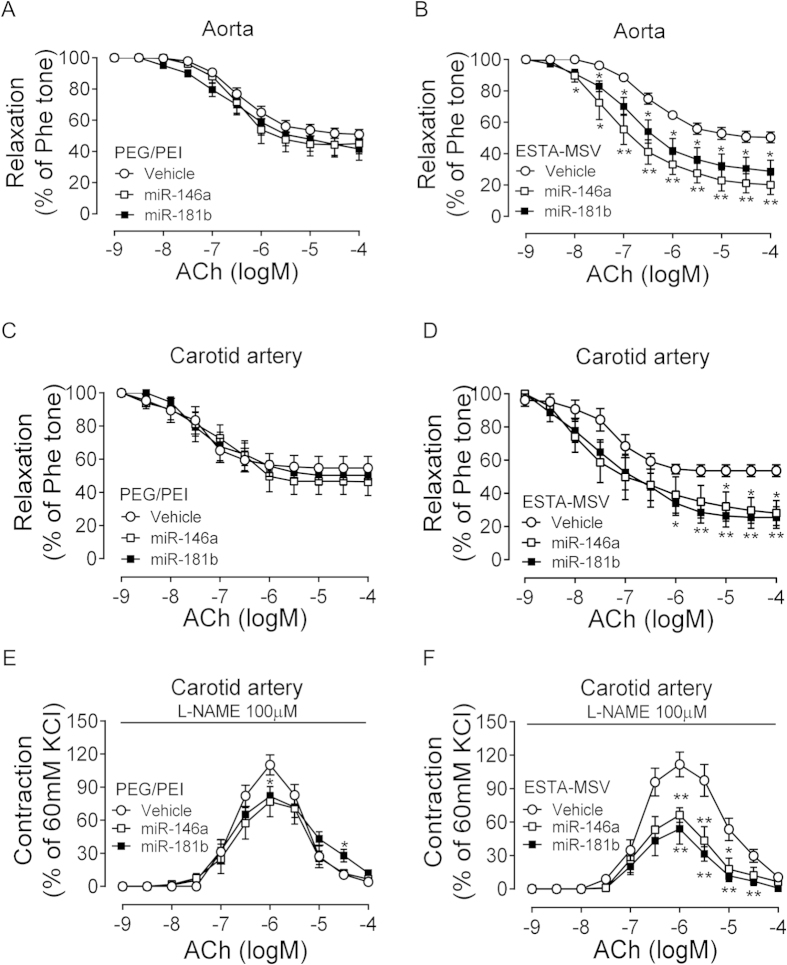
Figure 3. Effects of PEG/PEI/miRs and ESTA-MSV/miRs on the endothelial function of ApoE−/− mice.
The ACh-induced relaxation of Phe-precontracted abdominal aortas of ApoE−/− mice after intravenously injected with vehicle, miR-146a, and miR-181b (15 μg) loaded in PEG/PEI nanoparticles (A) or ESTA-MSV microparticles (B) biweekly for 12 weeks. (C,D) The ACh-induced relaxation of carotid arteries of ApoE−/− mice treated as above. (E,F) The ACh-induced contraction of carotid arteries of ApoE−/− mice in the presence of L-NAME (100 μmol/L). Data are shown as the means ± SEM (n = 5). *P < 0.05, **P < 0.01 vs. vehicle group.

