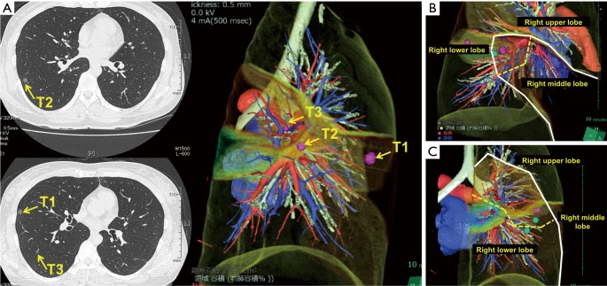
Figure 2.
Chest CT of a 30-year-old woman with three small pulmonary lesions (A). Preoperative marking of each lesion was performed using 3D-CT simulation. Then, wedge resection of the right middle lobe (B) and extended segmentectomy of the right lower lobe (C) were simulated preoperatively and finally performed with confidence. Pathological diagnosis revealed that all lesions were primary lung cancers. The dashed line indicates the surgical resection line. CT, computed tomography; 3D-CT, three-dimensional CT.

