
Figure 4.
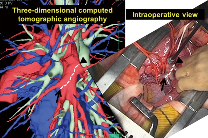
Comparison of the interlobar left pulmonary artery shown by 3D-CT and as seen in the operative view. Branches of the interlobar left pulmonary artery depicted by 3D-CT angiography were consistent with those seen in the actual operative field. 3D-CT, three-dimensional computed tomography.