
Figure 5.
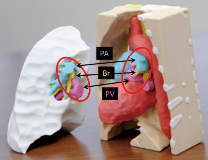
3D models of the RLL donor graft and post-pneumonectomy left thorax of the patient. These models were created accurately based on individual CT data. The PA, Br, and PV aligned in the same order in the donor and recipient (red circle). 3D, three-dimensional; RLL, right lower lobe; CT, computed tomography; PA, pulmonary artery; Br, bronchus; PV, pulmonary vein.