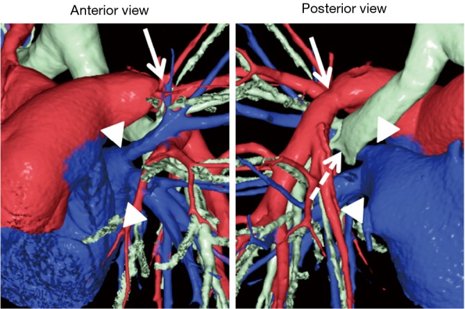
Figure 6.

Postoperative 3D-CT images in a bilateral t LDLLT with implantation of the inverted RLL in the left thorax. In this case, the donor pulmonary vein was sutured to the recipient’s left upper pulmonary vein as is usually performed in LDLLT (arrowheads). There were no complications involving the pulmonary arterial (arrows) and bronchial anastomoses (dashed arrow). 3D-CT, three-dimensional computed tomography; LDLLT, living-donor lobar lung transplantation; RLL, right lower lobe.
