
Figure 3. Evaluation of the metabolic activity of mammalian cells cultivated in the presence of silver.
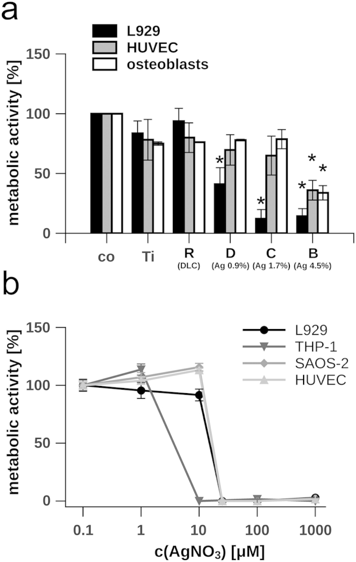
(a) L929 cells, primary osteoblasts and HUVEC were cultivated on different implants, while the metabolic activity was measured 24 h after seeding. The metabolic activity of cells grown on tissue culture-treated polystyrene (Co) was normalised to 100%, background levels of absorbance were subtracted. The silver content of the DLC is indicated in percentage. (b) Dose-dependent effect of silver nitrate (concentrations range from 0.1 to 1000 μM AgNO3) on the metabolism of L929, THP-1, SAOS-2 and HUVEC. Metabolic activity of non-treated cells was normalised to 100%, background levels of absorbance were subtracted. Data are represented as mean ± SD of at least 3 independent experiments. Statistical significant differences (P < 0.01) to the control group (co) are indicated by an asterisk. Ti = Ti6Al4V alloy; R = Ti6Al4V alloy with diamond-like carbon (DLC); B = DLC with 4.5 ± 0.5% silver; C = DLC with 1.7 ± 0.4% silver; D = DLC with 0.9 ± 0.2% silver.