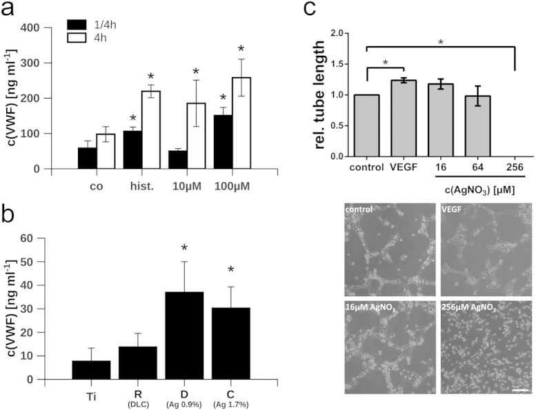
Figure 5. Analysis of VWF exocytosis.
(a) Release of von Willebrand factor (VWF) after treatment with silver nitrate (10 μM and 100 μM) for 15 min (black bars) or 4 h (white bars). Co = tissue culture-treated polystyrene; hist. = 50μM histamine. (b) Release of VWF from HUVEC grown on implants. VWF release was induced by stimulation with 50μM histamine as positive control. The silver content of the DLC surfaces is indicated in percentage. (c) Relative average tube length of HUVEC in the presence of different silver nitrate concentrations (AgNO3) and VEGF-A (50 ng/ml) as a positive control. Representative images showing the tube formation capacity of HUVEC are depicted. Scale bar corresponds to 100 μm. Data are represented as mean ± SD of at least 3 independent experiments. Statistical significant differences (P < 0.01) to the control group (co) or titanium surfaces (Ti) are indicated by an asterisk. Ti = Ti6Al4V alloy; R = Ti6Al4V alloy with diamond-like carbon (DLC); B = DLC with 4.5 ± 0.5% silver; C = DLC with 1.7 ± 0.4% silver; D = DLC with 0.9 ± 0.2% silver.

