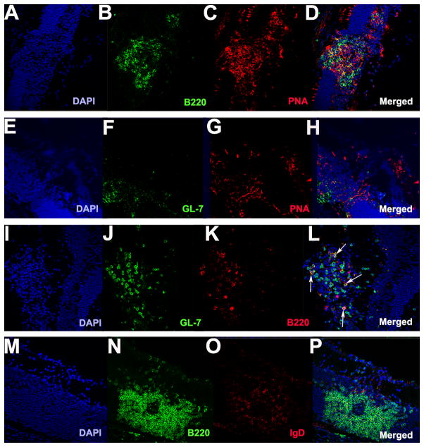
Figure 4. Retinal lymphoid aggregates stain positive for germinal markers PNA and GL-7 and low for IgD expression.
A–P. Retinal lymphoid aggregates. A–D. Retinal lymphoid aggregates with a distinct B cell zone area (B220 shown in green) and staining positive for the germinal center marker PNA (red) as shown in (C) under low magnification (20x). E–H. Retinal aggregates stain positive for GL-7 (green) and PNA (red) under low magnification (20x). I–L. Depicts the presence of GL-7+/B220+ cells in the retinal aggregates under high magnification (40x). White arrows shown in L denote GL-7+/B220+ cells. M–P. Illustrates low IgD (red) expression in the retinal lymphoid aggregates under high magnification (40x).

