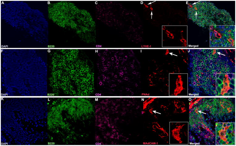
Figure 7. Lymphatic vessels and high endothelial venules are present in the retinal TLT.
A–O. A–E. Lyve-1 staining (red) showing lymphatic vessels (white arrows 40x), enlarged in the inset (63x, panel D). F–J. PNAd staining (red), and K–O MAdCAM-1 staining (red) showing HEVs (white arrows, 40x), enlarged in the inset (63x panels I and N). Note their “plump” appearance, compared to panel D, typical of HEVs. Lymphocytes are present in and around the HEVs (J, O). Each marker was visualized on separate sections of the same TLT aggregate to avoid artifacts due to possible cross-reactivity between secondary antibodies.

