
Figure 8. Retinal TLT stains positive for the stromal cell marker CD35 and follicular dendritic cell marker FDC-1, indicating the formation of organized networks within the retinal structures.
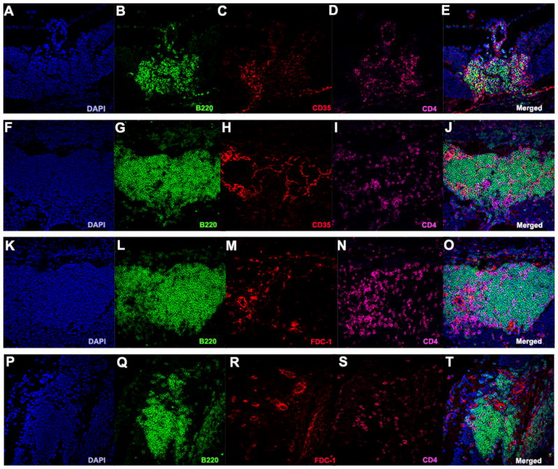
A–J. Retinal TLT stained positive for CD35 (red). K–T. Retinal TLT stained for FDC-1 (red) under high magnification (40x).