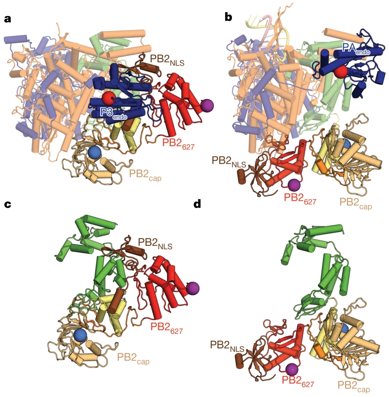
Figure 2. Comparison of apo-FluPolC with promoter-bound FluPolA.
a, Apo-FluPolC, depicted in the same orientation and colouring as in Fig. 1b, but with the C-terminal domains of PB2 coloured as in Fig. 1e. Domains that do not change between apo and promoter-bound conformations are depicted as semi-transparent. b, Promoter-bound FluPolA, shown as in a. c, d, The PB2 subunits of apo-FluPolC (c) and promoter-bound FluPolA (d), depicted as in a and b.

