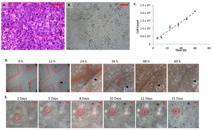
Figure 1.
Characteristics of mammary carcinoma (FMC2u) cells. (A) H&E of primary tumor isolated from late stage PyVT female at 40×, Scale bar = 50 µm; (B) Micrograph of FMC2u in cell culture at 20 Nguyen, ×, Scale bar = 20 µm; (C) Proliferation of FMC2u cells with an approximate doubling time of 24 h; (D) Migration assay. Red lines indicate a cross section “X” cut in the initial monolayer, approximately 1 mm in diameter. Cells were maintained in RPMI with 10% fetal bovine serum (FBS). Images taken every 12 h at 4×. Wound was completely closed by 60 h; (E) Colony formation in soft agar. 10,000 cells seeded on 0.8% agar RPMI and covered with 0.4% agar RPMI. Images were taken every 24 h at 10×. Purple circle follows the growth of a few cells at 48 h to a solid colony at 15 days. Scale bar = 100 µm.

