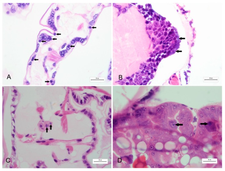
Figure 1.
Illustration of viral inclusion bodies in the histological sections of diseased M. rosenbergii larvae. Pale to dark basophilic, intracytoplasmic inclusion bodies with 2.8 to 4.0 μm in diameter are observed in a number of cells (indicated by arrows) in appendage epithelial tissues (A); ganglion (B); connective tissue (C); and hepatopancreas (D). All H and E. Bars = 20 μm.

