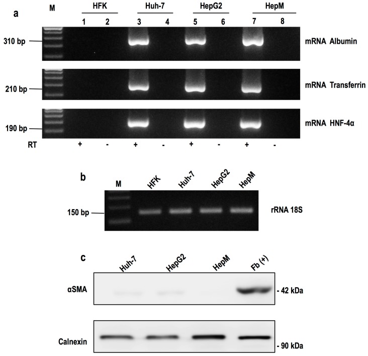
Figure 1.
Expression of hepatic markers by RT-PCR and Western blot: (a) Markers of hepatic mRNAs were amplified. Electrophoresis agarose seen in the amplified fragments of 310, 210 and 190 bp, corresponding to albumin, transferrin and HNF-4α, respectively. The fragment was generated from the cDNA obtained from total RNA Huh-7, HepG2 and HEPM cells. Huh-7 and HepG2 cells were used as positive control. HFK cells were used as negative control. Reverse transcriptase in the absence of product is not detected (lanes 2, 4, 6, 8). M corresponds to the marking of 100 bp molecular size; (b) Constitutively expressed gene 18S rRNA was used as an internal control; (c) Expression of the protein αSMA. By Western blot, the protein expression of αSMA was determined in the hepatic (Huh-7, HepG2 and HepM) and fibroblast cells (Fb (+)). Constitutively expressed calnexin was used as an internal control.

