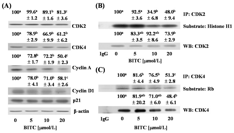
Figure 4.
BITC reduces the expression of cyclin A and cyclin D1 and inhibits the activity of CDK2 and CDK4 in TRAMP-C2 mouse prostate cancer cells. Cells were plated in 100 mm dishes at 1 × 10⁶ cells/dish. Twenty-four h after plating, the monolayers were serum-deprived in serum-deprivation medium for 6 h. Cells were then incubated for 3 h with serum-deprivation medium containing various concentrations of BITC (0, 5, 10, or 20 µmol/L); (A) The expression of CDK2, CDK4, cyclin A, and cyclin D1 were estimated by immunoblotting. The relative intensity of each band (normalized with its own β-actin) is shown above each band; (B,C) Cell lysates were incubated with an anti-CDK2 (B) or an anti-CDK4 (C) antibody and the immune complexes were precipitated with protein A sepharose. For the in vitro kinase assay, immunoprecipitated proteins were incubated with a substrate (B: Histone H1, C: retinoblastoma protein (Rb)) and [γ-32P]ATP. Each sample was subjected to SDS-PAGE and the gel was dried. The bands were visualized by autoradiography. For Western blot analysis, immunoprecipitated proteins were analyzed by Western blotting with an anti-CDK2 (B) or an anti-CDK4 (C) antibody. The relative intensity of each band was quantified and the control (0 μmol/L BITC) levels were set at 100%. Data denotes the mean ± SEM (n = 3). Means without a common letter differ, p < 0.05.

