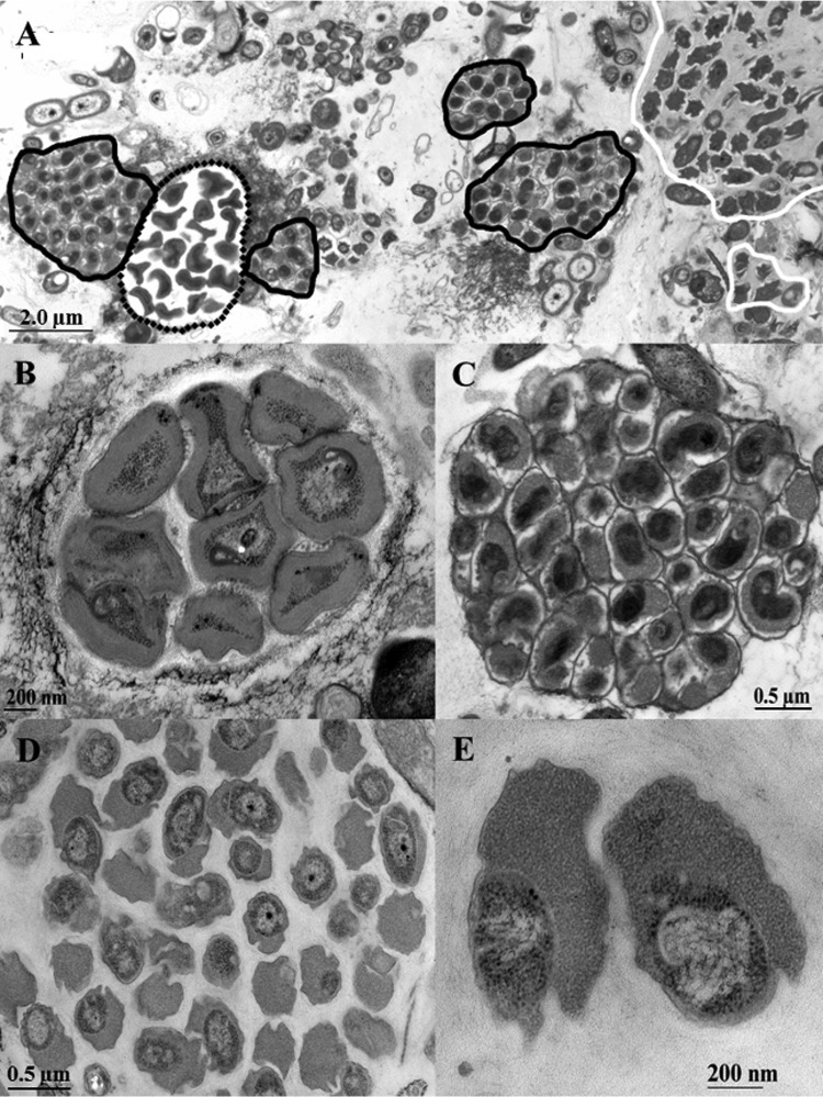
FIG 1.
Ultrathin section of the nitrifying biofilm grown on a carrier element of BF1 sampled in July 2010 and visualized via TEM. (A) Overview of the nitrifier arrangement inside the biofilm. Microcolonies of AOB Nitrosomonas are marked with dotted lines; for the second step of nitrification, Nitrospira (black outline) and Nitrotoga (white outline) were present. (B to D) Microcolonies of Nitrosomonas (B), Nitrospira (C), and Nitrotoga (D) in detail. (E) Typical ultrastructure of Nitrotoga cells, revealing the extremely wide periplasmic space with a particulate appearance.

