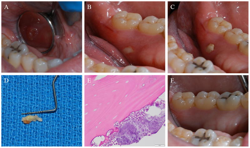
Fig. 1.

Clinical picture at initial presentation showing a gingival swelling (A), clinical picture a month later showing a 2 × 2 mm area of exposed bone (B), clinical picture 3 weeks later showing a 10 × 5 mm area of exposed necrotic bone (C), specimen removed and submitted for histopathologic evaluation (D), photomicrograph H&E (×200) showing a non-vital bone (sequestrum) with bacterial colonies (E), and a clinical picture of the healed area at 2 month follow-up examination (F).
