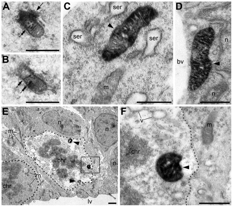
Fig. 2.

Anti-CB1–L31 made-in-Guinea pig antibody detected two types of immunopositive mitochondria in the neocortex of E13.5 mouse embryos: type 1 (A, B) and type 2 mitochondria (C-F). (A, B) Serial micrographs of a type 1 mitochondrion in the marginal zone. Notice characteristic immunoprecipitation in the cristae (double arrows) and around the mitochondrion (arrow). (C, D) Type 2 mitochondria (arrowheads) in an immature projection neuron (C) and an endothelial cell (D). Notice robust staining in the mitochondrial matrix whereas cristae are immunonegative. (E, F) Two mitotic metaphase cells (outlined with the dotted lines) in the ventricular zone facing the lateral ventricle (lv). The cell in the left lower corner of E contains only immunonegative mitochondria (m) and no detectable ultrastructural pathologies. In contrast, the adjacent mitotic cell (in the center of E) contains multiple type 2 mitochondria (arrowheads) and evidences for degradation, as shown by numerous empty vacuoles (v) and diminished electron density in the cytoplasm. The framed area in E is enlarged in F. Scale bars: 1 μm (E); 0.5 μm (A-D, F). bv, lumen of blood vessel; chr, chromosomes in mitotic cells; n, cell nucleus; ser, swollen cisterns of rough endoplasmic reticulum.
