Fig. 6.

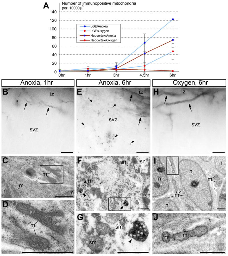
Oxygenation of postmortem E13.5 mouse embryonic brains attenuates the rise of immunopositive mitochondria (detected with CB1–L31 made-in-Guinea pig serum) and necrosis-like ultrastructural pathology. (A) Quantification of immunopositive mitochondria in the subventricular zone (svz) of postmortem brains in anoxia conditions or with oxygenation. Shown are the average ± SD. (B-J) Representative light (B, E and H) and electron micrographs (C, D, F, G, I, and J) of anti-CB1 labeling of the neocortical SVZ and intermediate zone (iz) that contain normal CB1-positive axons (arrows in B, E and H) under the indicated conditions. (B-D) At 1 hr anoxia, postmortem neurons retain normal structure of nuclei (n) and organelles including mitochondria (m) that are negative for anti-CB1–L31 labeling. (E-G) At 6 hr anoxia, postmortem neurons show numerous immunopositive mitochondria (arrowheads) and necrosis-like ultrastructural pathologies, such as spherical-shape nuclei (sn) and swollen mitochondria (sm). (H-J) In contrast, dissected embryonic brains immersed in the oxygenated medium demonstrate very few, if any, immunopositive mitochondria (H) and mostly normal ultrastructure in postmortem neurons (I and J). Framed areas in C, F and I are enlarged in D, G and J, respectively. Scale bars: 10 μm (B, E and H); 1 μm (C, D, F, G, I, and J). m, mitochondria; n, nucleus; svz, subventricular zone.
