Figure 5.

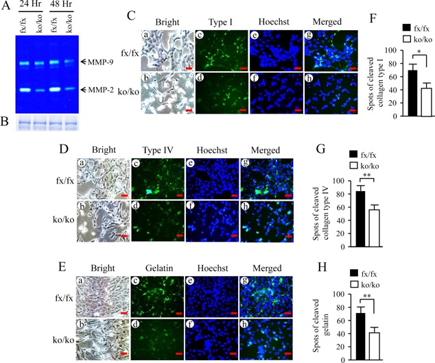
Deletion of BMP2/4 genes leads to impairment of extracellular matrix remodeling. (A) The equal amount of supernatant collected from the iBMP2/4fx/fx and iBMP2/4ko/ko cells was analyzed using gelatinolytic activities. Expression of Mmp‐2 and Mmp‐9 in the iBMP2/4ko/ko cells was decreased. (B) The equal amount of supernatant harvested from the iBMP2/4fx/fx and iBMP2/4ko/ko cells was loaded onto a 10% SDS‐PAGE gel and stained by Coomassie brilliant blue dye. (C–E) The iBMP2/4fx/fx and iBMP2/4ko/ko ob cells were grown on the DQ‐FITC‐collagen types I‐, IV‐, and DQ‐FITC‐gelatin‐coated slides for 12 h. The cells were fixed and degraded spots of the DQ‐FITC‐collagen types I, IV, and DQ‐FITC‐gelatin were observed using Nikon inverted fluorescent microscope (c and d). (a and b) The cells were photographed under a light inverted microscope. (e and f) The cells were treated with Hoechst dye for nuclei staining. (g and h) The images were merged. (F–H) Spots of the cleaved collagen type I, IV, and gelatin were quantitated from the iBMP2/4fx/fx and iBMP2/4ko/ko ob cells. There are significant differences between the iBMP2/4fx/fx and iBMP2/4ko/ko ob cells. *P < 0.05; **P < 0.01. Scale bars, 20 µm. fx, floxed; ko, BMP2/4 knock‐out.
