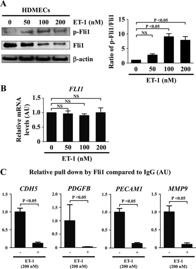
Figure 1.
Effect of endothelin 1 (ET-1) on Fli-1 expression and function in human dermal microvascular endothelial cells (HDMECs). A, HDMECs were treated for 24 hours with ET-1 at the indicated concentrations, and the expression and phosphorylation levels of Fli-1 were determined by immunoblotting with whole cell lysates (β-actin was used as a control for equal loading). The fold change in the ratio of phospho–Fli-1 to total Fli-1 (quantified by densitometry) was determined. B, Levels of mRNA for Fli-1 after the same treatment as described in A were evaluated by quantitative reverse transcription–polymerase chain reaction (qRT-PCR). C, HDMECs were treated with ET-1 (200 nM) or vehicle for 2 hours and analyzed by chromatin immunoprecipitation. The binding of Fli-1 to the target gene promoters (CDH5, PDGFB, PECAM1, and MMP9) in response to ET-1 as compared to unstimulated controls (set at 1) was quantified by qRT-PCR. Values are the mean ± SEM (n = 3 mice per group). NS = not significant.

