Fig. 1.

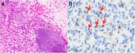
PMT with TIO. a: The tumor was characterized by the proliferation of bland spindle or oval cells, with grungy calcification. b: By immunohistochemical staining, FG322-3 expression showed a distinct, dot-like staining pattern (arrows)

PMT with TIO. a: The tumor was characterized by the proliferation of bland spindle or oval cells, with grungy calcification. b: By immunohistochemical staining, FG322-3 expression showed a distinct, dot-like staining pattern (arrows)