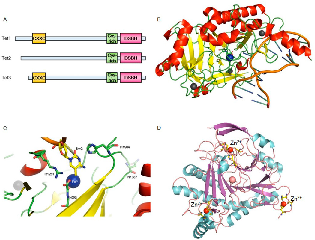
Figure 3.
Overall domain architecture of TET proteins and the published human TET2 structure. (A) TET proteins contain a CXXC DNA-binding domain and a conserved catalytic domain composed of a Cys-rich region and a DSBH fold. (B) Crystal structure of the TET2–DNA complex. The Cys-rich regions bind zinc cations and are folded together with the catalytic domain. (C) Close-up view of the active site of TET2. DNA is colored orange; 5mC, NOG, and critical residues are shown in stick representations, iron is shown in a blue sphere, and zinc is shown in a gray sphere. (D) Ribbon representations of three zinc cations in TET2. Zinc cations are shown and labeled in red spheres; corresponding coordination residues are shown in stick representations.

