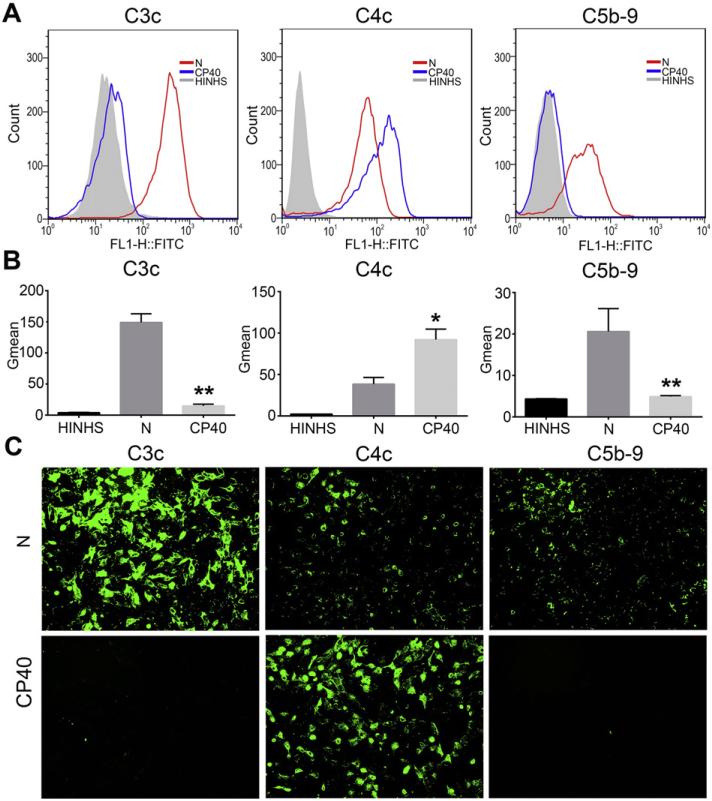
Fig. 3.
Cp40 significantly inhibits the cleavage of complement C3 and the formation of C5b-9. Deposition of C3c- and C4c-containing complement opsonins and C5b-9 on iPECs was detected by FACS (A and B) and immunofluorescent staining (C, ×100) after the incubation of iPECs with either 20% NHS (N) or Cp40-pretreated 20% NHS (Cp40). Cells incubated with HINHS were used as negative controls. For FACS analysis, the Gmean was used to evaluate the degree of complement deposition. (A) FACS results shown are representative of three independent experiments. (B) The degree of deposition of C3c- and C4c-containing opsonins, and C5b-9 on iPECs is shown in the bar graphs. Data shown are means ± SEM (*P < 0.05, **P < 0.01 vs. the N group; n = 3 per group). (C) Images are representative of three independent experiments.

