
Figure 2. PTPδ and TrkB compete for binding to Slitrk5.
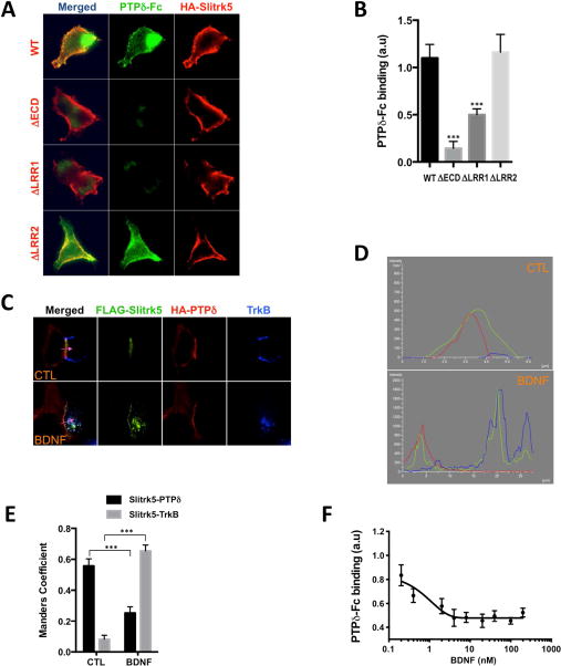
(A) Slitrk5 binds PTPδ through LRR1. Soluble purified PTPδ-Fc chimeras were added to HEK293T cells expressing WT and indicated deletion mutants of Slitrk5 and the binding analyzed by immunofluorescence microscopy. Note that the observed binding to WT Slitrk5 is abolished by deletion of Slitrk5’s extracellular domain (ECD) or LRR1 but not LRR2. (B) Quantitative analysis of these results. Results are means ± SEM from 3 independent experiments. 20–30 cells were analyzed per condition per experiment. ***P< 0.0001 significantly different from WT condition. (One-way ANOVA and Dunnett’s multiple comparisons test). (C) BDNF displaces Slitrk5 binding from PTPδ to TrkB. Heterophilic cell adhesion assay in which HEK293-TrkB cells expressing FLAG-Slitrk5 are co-cultured with HEK293T cells expressing HA-PTPδ. Surface proteins were visualized by fluorescence microscopy in the presence or absence of BDNF treatment (as described in Supplemental Experimental Procedures). (D) Fluorescence intensity trace of FLAG-Slitrk5 (green), HA-PTPδ (red), and TrkB (blue) scanning across corresponding purple arrow as indicated in (C). (E) Quantitative colocalization analysis of FLAG-Slitrk5-HA-PTPδ and FLAG-Slitrk5-TrkB in the presence or absence of BDNF treatment in (C). Results are means ± SEM from 3 independent experiments. 10–15 cells showing heterophilic adhesion were analyzed per condition per experiment. ***P< 0.0001 significantly different from control condition. (2-way ANOVA and Sidak’s multiple comparisons test). (F) BDNF-induced dissociation of pre-bound PTPδ-Fc from HA-Slitrk5-expressing HEK293-TrkB cells. HA-Slitrk5-expressing HEK293-TrkB cells were pre-incubated with saturating condition of PTPδ-Fc (400 nM) for 1hr. After washing, cells were incubated with indicated dose of BDNF for 30 min. Remaining PTPδ-Fc binding was analyzed by immunofluorescence microscopy. Results are means ± SEM from 3 independent experiments. 20–30 cells were analyzed per condition per experiment.