
Figure 6. Slitrk5 interacts with Rab11-FIP3 to facilitate TrkB receptor trafficking to Rab11-positive recycling endosomes.
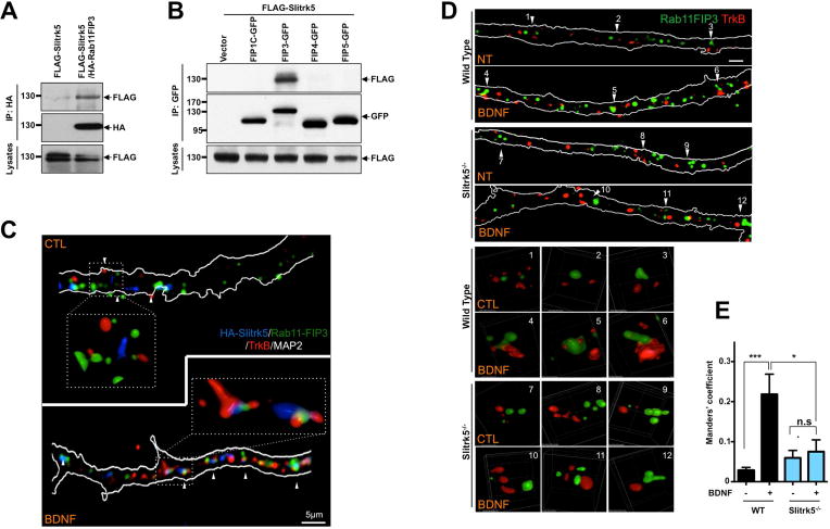
(A) and (B). Representative blots showing specific interaction between Slitrk5 and Rab11-FIP3. (A) HEK293T cells were transfected with cDNAs encoding FLAG-Slitrk5, and HA-Rab11-FIP3. Cell lysates were immunoprecipitated with anti-HA antibodies and immunoblotted with anti-FLAG antibodies. (B) HEK293T cells were transfected with cDNAs encoding FLAG-Slitrk5, and either empty vector, Rab11-FIP3-GFP, FIP1C-GFP, FIP4-GFP or FIP5-GFP. Cell lysates were immunoprecipitated with anti-GFP antibodies and blotted with anti-FLAG antibodies. (C) Representative image showing the co-localization of TrkB and Slitrk5 with Rab11-FIP3 with BDNF-dependent manner. WT striatal neurons expressing HA-Slitrk5 were stimulated with or without BDNF after incubating with anti-TrkB and anti-HA antibody for surface protein labeling. Neurons were stained with anti-Rab11-FIP3 antibody after fixation and permeablization. Super resolution images were acquired using a Nikon N-SIM structured illumination microscope. (D) Representative images showing requirement of Slitrk5 for TrkB receptor localization in Rab11-FIP3 compartments. Co-localization of TrkB and Rab11-FIP3 was examined with WT and Slitrk5−/− striatal neurons in the presence or absence of BDNF. Lower panels showed enlarged images of numbered regions in upper panels. (E) Co-localization of TrkB and Rab11-FIP3 was quantitated as described in Experimental Procedure. Results are presented as means ± SEM from 3 independent experiments determined from analysis of n ≥ 20 neurons per condition per experiment (*P<0.05, ***P<0.001, Student’s t test).