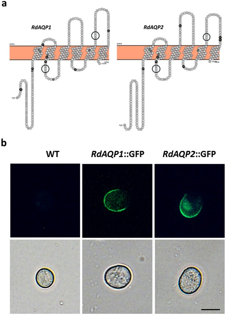
Fig 5. Predicted topology and localization of RdAQP1 and RdAQP2 proteins.
(a) Hypothetical prediction of RdAQP1 and RdAQP2 topology by the TMHMM method, drawn by the Protter-visualize proteoforms program [53], showing the proteins' six transmembrane domains. The highly conserved NPA domains are circled. The Cys and His amino acids are filled in gray and black color, respectively. (b) Expression of RdAQP1::GFP and RdAQP2::GFP in R. delemar spores. Bar = 10 μm.

